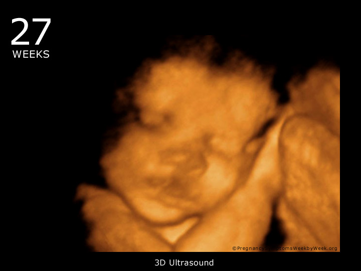

The length of your baby can be measured on an ultrasound by measuring the distance between your baby's head (crown) and his bottom (rump). The head is still about half the total length of your baby. 12(3):28-31.Your baby takes on a more human form as his neck lengthens and his head is seen as separate from his body. Australian Journal of Ultrasound in Medicine. 3D ultrasound in first and second trimester - hype or helpful?. Avoid Fetal "Keepsake" Images, Heartbeat Monitors. The American College of Obstetricians and Gynecologists.

Guidelines for Diagnostic Imaging During Pregnancy and Lactation.

Ultrasound for fetal assessment in early pregnancy. Learn more about our editorial and medical review policies. We believe you should always know the source of the information you're seeing.
#4d ultrasound video at 10 weeks professional#
When creating and updating content, we rely on credible sources: respected health organizations, professional groups of doctors and other experts, and published studies in peer-reviewed journals. That's why most healthcare providers don't use 3D ultrasound regularly.īab圜enter's editorial team is committed to providing the most helpful and trustworthy pregnancy and parenting information in the world. For most pregnancies, 3D ultrasound won't give any more usable information than a standard 2D image. These early scans are used to date a pregnancy and due date, and check on suspected problems such as ectopic pregnancy.įind out more about ultrasounds during pregnancy. If your provider needs to do an ultrasound in the first trimester, she may use a vaginal probe to get closer to your uterus. Many women will have an earlier 2D ultrasound as well. When you're done, the technician will probably give you a few black-and-white images as a keepsake. You'll be able to hear the heartbeat and if your baby is awake, you'll see movement on the screen. These waves bounce off your baby, and a computer translates the echoing sounds into video images that reveal details of your baby's body, position, and movements. During your ultrasound, a technician will use a handheld instrument called a transducer to send sound waves through your uterus. You'll probably have a 2D ultrasound about halfway through your pregnancy (between 18 and 22 weeks). What ultrasounds will I have during pregnancy?
#4d ultrasound video at 10 weeks series#
In a 4D ultrasound, a series of 3D images is put together to form a low-resolution video. 4D ultrasound adds a fourth dimension – time.3D ultrasound uses the same basic idea as 2D ultrasound, but takes many images from different angles and processes them together to create an image that looks like a real photograph.This process creates simple, black-and-white images that create a cross-section view, with bright spots for denser materials like bone. 2D ultrasound is the standard ultrasound that healthcare providers use.

The waves bounce off tissues to create a picture on a screen. The ultrasound machines used for medical imaging use waves between 2 to 20 megahertz – that's about 100 times higher than the top of the range we're able to hear (20 to 20,000 hertz). It works just like the sonar on boats, which use sound waves to locate things underwater. Ultrasound is a way to look inside the body with high-frequency sound waves.


 0 kommentar(er)
